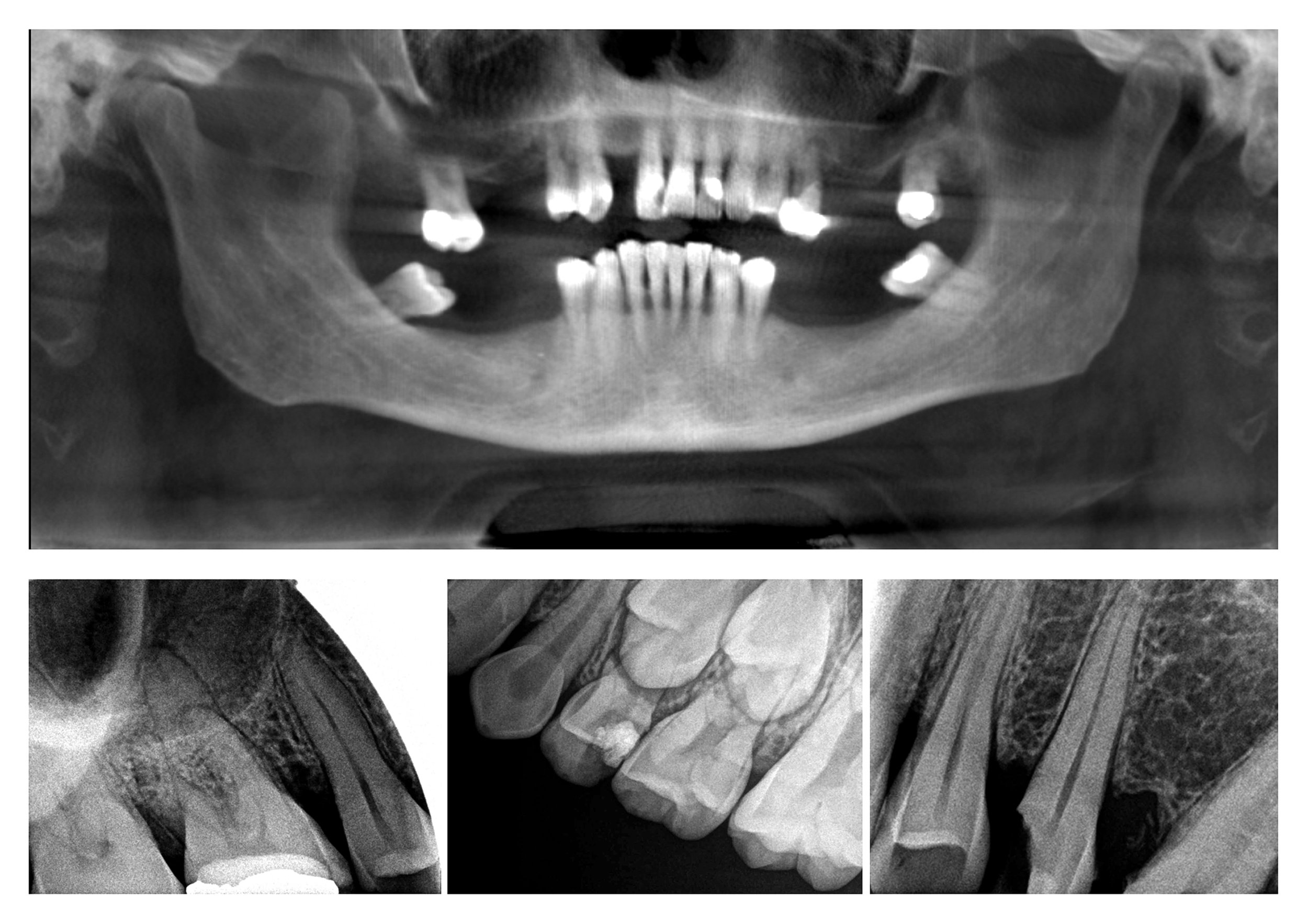
Digital Radiograph (X-rays)
How much radiation is involved in dental x-rays?
Dental x-rays are essential diagnostic tools and a necessary component for many dental procedures and for identifying a number of diseases within the oral cavity. In our office, we use an electronic sensor instead of x-ray film to capture and store the digital images on a computer. The digital image can be manipulated or enlarged to help the dentist detect problems easier.
Digital x-rays reduce radiation exposure by up to 90% of traditional film x-rays. A routine exam (4 bitewing x-rays) can be measured to be approximately 0.005 mSv, which is about the amount of radiation exposure from a short airplane flight. However, the dentist (knowing the health of the patient and their vulnerability to oral disease) is in the best position to weigh the benefits of taking dental radiographs against the risk of exposing a patient to X-rays.
Are dental x-rays safe?
Even though digital x-rays produce a low level of radiation and are considered very safe, dentists still take necessary precautions to limit the patient's exposure to radiation. Only necessary x-rays are taken and lead aprons and thyroid collars are used to shield the patient's body.
How often should dental x-rays be taken?
The need for x-rays depends on each individual's risks and health needs. The dentist will recommend necessary x-rays based upon the medical and dental history of the patient, signs and symptoms.
Full mouth series of x-rays is generally considered good for diagnostic use every 3-5 years. Bitewing x-rays done at a recall exam have a general diagnostic life of 1 year. However, x-rays are not prescribed at every visit and dentists make an effort to limit the amount of radiation exposure to all patients.
What is a dentist looking for on an x-ray?
Dental x-rays may reveal:
- Dental decay (in the enamel and dentin layers)
- Gum disease (changes in bone levels)
- Abscess or cysts (dental infection)
- Cancerous or Benign tumors of hard tissue
- Developmental abnormalities (ie. missing permanent teeth)
- Tooth and root positions
- Sinus and nerve canal positions
- Quantity and Quality of bone
- Sources of infection
- Sources of pain
- Signs of systemic disease
If a dentist cannot accurately determine the proper information from a two-dimensional x-ray, a Panoramic or Cone Beam (CBCT) image may be prescribed.

